Research

Using Dictyostelium, we identified a pathway in which the dimeric acidic protein 14-3-3 acts genetically between microtubules, racE and myosin II to control cell shape, cortical tension, and cytokinesis.  In this context, 14-3-3 requires GTP and racE for cortical association, controls the extent of microtubule-cortex interactions, and binds myosin II heavy chain, controlling its assembly into bipolar thick filaments.  This first study was simplified by using Dictyostelium cells, which only has one myosin II and one 14-3-3 protein and was presented in Zhou et al. Curr. Biol. 2010.

Humans have three nonmuscle myosin II and seven 14-3-3 proteins.  We expressed and purified all 10 proteins and discovered that the 14-3-3s can also modulate human nonmuscle myosin II assembly, particularly myosin IIB.  The study of the human proteins was presented in West-Foyle et al. 2018.

Read More

For decades, pathologists have known that formation of cell-in-cell structures, where one cell engulfs another (entosis), could be frequently observed in human tumors.  We found that these structures require cell-cell adhesion and a mechanical differential between the inner and outer cells.  Specifically, the inner cell has ~2-fold greater elasticity than the outer cell.  This mechanical differential comes from inactivation of myosin II in the outer cell and activation in the inner cell.  Major human oncogenes such as activated KRAS are sufficient to create the mechanical differential by down-regulating Rho pathways that normally activate myosin II.  At least in culture, this mechanical heterogeneity facilitates cell-cell competition where the inner (stiffer) cell loses and the outer (softer) cell wins.  For more information, please see Sun et al. Cell Res. 2014 and Hamann et al. Cell Rep. 2017.

Read More

Over many years, we have deciphered the mechanochemical system that governs cytokinesis cell-shape change.  While the textbook view has the mitotic spindle as the principle driver, the system is really structured as a control system with many feedback loops.  The long axis rule, which was described in the 19th Century and states that the mitotic spindle will elongate along the long axis of the cell, already predicts such a feedback system.  Indeed, the mitotic spindle sends cues to the equatorial and polar cortices.  These cues help drive regional accumulation of equatorial and polar proteins that really exist as inverse concentration gradients across the cortex.  The polar cortex proteins then define the regional viscoelasticity and resistive stresses against which the contractile equator must work.  The polar cortex also generates expansive stresses, which help elongate the cell but primarily requires cell adhesion to be effective.  The equatorial system defines the regional viscoelasticity of the cleavage furrow, generates contractile stresses, and is highly mechanoresponsive (senses mechanical stresses and tunes protein concentration and dynamics as a result).  This mechanosensitivity endows the cell with a tremendous dynamic range of force production with about 5-fold coming from myosin II’s load sensitivity (reflected in changes in duty ratio) and another 5-7-fold coming from changes in protein concentration.  Overall, the system has a 30-50-fold dynamic range in force-generating ability.  The bases for these concepts may be found in Zhang and Robinson, PNAS 2005; Reichl et al. Curr. Biol. 2008; Poirier et al. PLoS Comp. Biol. 2012; and Kee et al. Mol. Biol. Cell 2012.

Read More

We are identifying new, critical biology underlying airway diseases, such as chronic obstructive pulmonary disease and other fibrotic lung diseases.  To do this, we have built a platform, which leverages a model system, Dictyostelium discoideum, to identify relevant genes  for protecting cells from toxins, such as cigarette smoke.  We find that these genes are also protective in primary human airway epithelial cells and protect several key functions such as airway surface hydration and ciliary function.  The unexpected biology that we have uncovered is paving the way for small molecule screens, which we hope will facilitate the discovery of new therapeutic strategies for these currently incurable lung diseases.

Check out our Kliment et al. 2021 publication where we describe the identification of adenine nucleotide translocase (ANT) as a protector from cigarette smoke in Dictyostelium and in human primary airway epithelia. We also discovered that smokers and patients with COPD have reduced ANT expression.  This work was featured in Scientific American.

Read More

While much is understood about how myosin II is regulated by light chain and heavy chain phosphorylation, we still do not know how the protein is specifically localized.  Mechanosensing through cooperative binding of myosin motors to allosteric actin filaments is one major mechanism.  To identify new mechanisms, we performed genetic suppression of a phosphomimic (3xAsp) myosin II, which poorly assembles into bipolar thick filaments, and found several genetic suppressors that rescued assembly and/or cleavage furrow accumulation.  Several new proteins were identified, including the Regulator of Microtubule Dynamics-1 and methylmalonate semialdehyde dehydrogenase.  For more information, please see Ren et al. 2014.

Read More

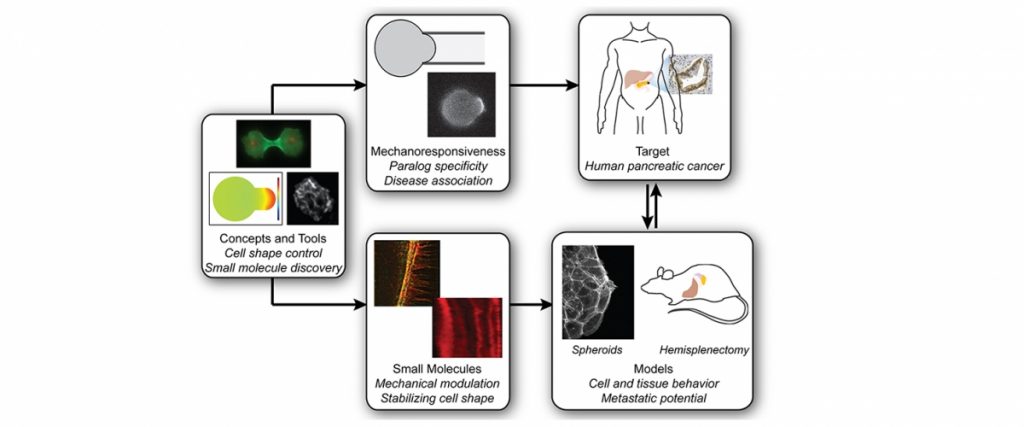
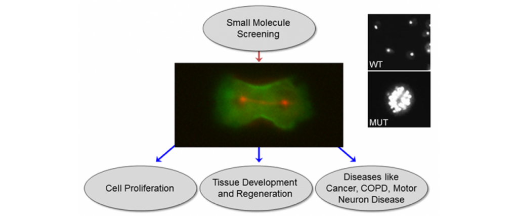
We have created a platform-based approach to small molecule discovery based on the concept that major health challenges, namely cancer metastasis and chronic obstructive pulmonary disease, are associated with defects in control of cell and tissue morphology and mechanics. We bring a unique suite of tools, drug screening strategies, and expertise for identifying and developing novel chemical modulators (future drugs) of cell morphology to attack these major health problems.  Most strategies for drug discovery draw upon small molecule screening against specific target proteins. However, these compounds often lead to effects at the cell and organism level that cannot be anticipated. Instead, we recognize that diseases are ultimately manifested in alterations in cell and tissue behaviors. These behaviors are controlled by networks of biomolecules, not any single one. Therefore, we start our drug discovery approaches by identifying small molecules or combinations of molecules that directly modulate these cell and tissue behaviors and then use a variety of genetic, biochemical and biophysical approaches to identify the molecular bases for the compounds’ action. The combination of strategies constitute Biology-In-Action (BIA),  which is used to identify our compounds, and then Quantitative Phenotypic Fingerprinting (QPF), which is used to identify the targeted pathways and targets of the compounds.  Here we provide a summary of our work in just one of these areas, which is in oncology.

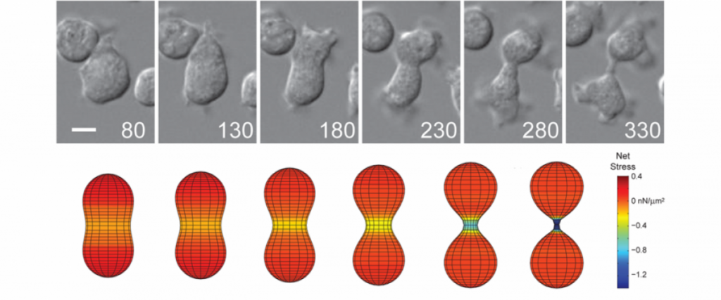
Importantly, for pancreatic cancer (an immediate focus of our work), the major drivers include activated mutant versions of an important signaling protein KRAS, inactive mutant forms of a quality control surveillance protein TP53, as well as a collection of other mutant proteins and proteins with significantly upregulated expression.   However, we found that many key proteins (e.g. specific versions of the motor protein myosin II, myosin IIA and IIC) in the cell’s powertrain are significantly upregulated.  Even more provocative, myosin IIA can slow and block tumor progression (tumor suppressive function).  This motor protein does this by modulating the power output and mechanical state (how stiff or tense the cell is).  Through these activities, myosin II activity also feeds back onto quality control proteins like TP53 (much like the speed of the car is fed back to the cruise control system so that engine power is modified to maintain course).  However, in cancer, many of the signaling pathways become altered leading to disrupted myosin II motor function.  When these systems become disturbed, the cell system is poised to grow uncontrollably, change shape, invade surrounding tissue, and even break free so that the cell or small clusters of cells can move to distant locations and set up new colonies (i.e. metastasize).

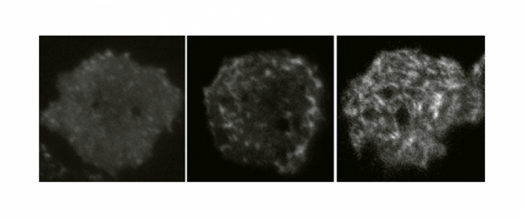
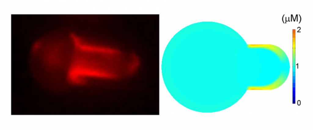
With our discovery platform, we have identified a strategy to activate the function of one myosin II form (myosin IIC), which is specifically overexpressed in pancreatic cancer. Using a small molecule (4-HAP), we can increase the amount of functional myosin IIC and correct many of the mechanical defects associated with pancreatic cancer progression. The consequence for the cancer is that the cells have reduced ability to migrate through obstacles, and in animal studies, 4-HAP reduces the ability of the cells to form metastases (Fig. 1). We also hypothesize that in addition to the reduction in the ability of the cancer cells to invade and metastasize, we are pushing the cells back towards a less cancerous state. This collective effect of targeting the powertrain to block metastasis and return cancer cell identity to a more normal state is a new innovative approach to treating cancer.

For more information on this work, please see Surcel et al. PNAS 2015, Surcel and Robinson PNAS 2019, Surcel et al. Cancer Research 2019 , Bryan et al. PNAS 2020 and Parajón et al. AJP Cell Physiol. 2020.

Read More

Chemical signal transduction cascades are often thought to be the principle drivers of protein localization.  However, we have found that mechanical stress is just as important and in many cases over-rides chemical cues.  Many cytoskeletal proteins, including myosin II, cortexillin I, IQGAP scaffolding proteins, and several others, lock in in the cytoskeletal network.  By having mechanosensation trumping other inputs, the cell has the intrinsic ability to build its cytoskeletal networks exactly where they are needed and then allow them to disassemble when their job is complete – i.e. the ultimate smart material.  Please see Srivastava and Robinson, Curr. Biol. 2015 for more information.

Read More

Cytokinesis is inherently a mechanical process, driven by active expansive and contractile stresses and Laplace pressure differentials (cortical tension x local curvature), all of which act upon the cell’s viscoelastic material.  By developing a complete mechanical description of the cell, we could account quantitatively for cytokinesis shape change, including the kinetics of furrow ingression.  Thus, cells divide by mechanics, not biochemistry.  The relevant question then becomes how does the cell use biochemistry to generate these mechanics.  The major papers describing this work include Zhang and Robinson, PNAS 2005; Reichl et al. Curr. Biol. 2008, Poirier et al. PLoS Comp. Biol. 2012., Srivastava et al. Curr. Biol. 2015, and Kothari et al. J. Cell Biol. 2019.

Read More

How can a cell build large macromolecular assemblies fast?  They do so by maintaining the critical components preassembled in the cytoplasm in the form of mechanoresponsive contractility kits (like IKEA kits).  Then, when the cell receives an input, such as a mechanical stress, the system responds by rapidly unpacking and building the network structures, allowing the cell to overcome or adapt to the input.  For a full explanation, please see Kothari et al. J. Cell Sci. 2019 (primary paper describing how these concepts and network were discerned) and Kothari et al. J. Cell Sci. 2019 (review describing cell shape control through the lens of control theory).

Read More

Among the earliest inputs that cells experienced, mechanical stress (forces) guide and direct behavior of cells, including when they are part of tissues, organs, and organ systems.  These mechanical stresses are propagated through the cell’s skin (the cell cortex), which is a composite material of membrane and cytoskeleton.  Key molecular machinery senses the forces, and through mechanotransduction, the mechanical signals may be converted into biochemical signals, which guide cell behavior.   Different proteins sense and respond to different types of deformation. For example, myosin II and alpha-actinin respond to dilation deformation while filamin responds to shear deformation.  For more information, please see Effler et al. Curr. Biol. 2006, Ren et al. Curr. Biol. 2009, Luo et al. Biophys. J. 2012, Luo et al. Nat. Mater. 2013, Schiffhauer et al. Curr. Biol. 2016, Schiffhauer and Robinson, Biophys. J. 2017, Thomas and Robinson Sem. Cell  Dev. Biol. 2017, and Schiffhauer et al. J. Cell Biol. 2019.

Read More

To decipher the relative contributions of the membrane and cortex to cells, we built a minimal cortex inside a giant unilamellar vesicle and compared the mechanics of the synthetic cell to those of wild type cells and cells where we progressively removed important cytoskeletal components.  By comparing the reconstituted cell with the ‘deconstituted’ cell, we found that the membrane contributed no more than 2% to total cell mechanics.  This maximum places important limitations on what membrane can contribute to cell mechanics.  For example, in cells where the membrane and cortex are tightly coupled, the cytoskeleton will bear nearly all of the load.  In cells that have loose coupling between the membrane and cortex, the membrane will be more sensitive to small stresses, though it still cannot bear much load without tearing.  For more details, please see Luo et al. App. Phys. Lett. 2014.

Read More

The central goal of our research is to discern fundamental principles of cell shape control and then to apply this knowledge to a variety of disease states.  Importantly, numerous diseases, including most cancers and lung diseases such as chronic obstructive pulmonary disease (COPD), derive a significant portion of their etiology from defects in cell mechanics.  Yet, cell mechanics has really not been explored as a source of novel therapeutic targets.  Because cell division showcases the cell’s mechanics at work and defects in cell division yield highly visible, easily scorable phenotypes (namely, multi-nucleation), we are using cytokinesis to identify small molecules that can tweak cell mechanics (Surcel et al. PNAS 2015).  We have reached the stage where our first small molecules have transitioned to animal studies of pancreatic ductal adenocarcinoma (PDAC) and colorectal cancer; see Surcel et al. Cancer Res. 2019 and Bryan et al. PNAS 2020.

Read More